| We are pleased to offer our patients the latest advancement in
Eye Care Technology! |
| |
 |
|
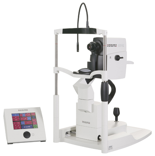
SPECTRALIS OCT |
|
Early recognition of disease helps to delay and prevent the most common causes of blindness. |
A SPECTRALIS® OCT examination provides information about the condition of the retinal layers. The examination can help identify early signs of disease in the retina and on the optic nerve head.
"OCT" is short for "Optical Coherence
Tomography". It is a modern imaging technique
like MRI or sonogram, which shows doctors
structures inside the eye that can change
due to eye disease.
In an OCT exam a light beam scans the eye
through the pupil. The beam scans across
the back of the eye, and the reflected light
is translated into a detailed image of the
structures within the retina.
OCT has become invaluable in advanced
eye care; because, it allows doctors to see
very tiny changes in the eye which would
otherwise be difficult to detect.
|
|
|
In an OCT exam a light beam scans the eye
through the pupil. The beam scans across
the back of the eye, and the reflected light
is translated into a detailed image of the
structures within the retina.
OCT has become invaluable in advanced
eye care; because, it allows doctors to see
very tiny changes in the eye which would
otherwise be difficult to detect.
Careful examination and analysis of the
structures seen in OCT images can help
us identify early signs of eye diseases
like glaucoma and AMD.
In fact, OCT is so sensitive, it often shows
us signs of disease before you notice
any changes in your vision. This is a tremendous
advantage to you; because, studies
have proven that starting treatment early is
the best way to save vision.
OCT is also helpful for confirming whether
your treatment is working or if alternate
treatments should be considered.
Preparation
At the start, you will be seated directly in front of the instrument. During the entire test, you will keep your head still with your chin resting on the chin rest and your head firmly against the instrument's head band. The examiner will sit opposite from you on the other side of the instrument. You always look directly into the instrument, staring at a fixation point or at the point indicated by the eyecare professional. You can blink normally. The examination takes just a short time
Examination
When the examination begins, a blue dot will appear in the instrument. You will be asked to stare at the blue dot during the entire examination. The examiner will move the instrument toward your eye without touching you. At a safe distance from the eye, the examiner is able to produce a good image of your retina or optic nerve head. A safe light beam scans the most important structures.
After examination
The examination is completely painless for you and is done without touching your eye. After the examination, your vision will not be impaired. If your eyecare professional has not dilated your pupil to conduct the OCT test or another examination, you can drive as usual.
|
| |
|
|
|
| |
|
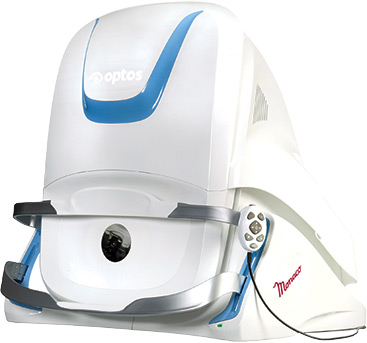
| OPTOS
Optomap |
We all want to to protect our eyesight and that is why it is important to have annual vision tests. This allows us to detect changes in the front of your eye so that alterations can be made to your eyeglass or contact lens prescription.. We also need to inspect the retina to check if it is healthy, damaged, or showing signs of disease.
When detected at early, many diseases can be treated without any further loss of vision. Each Optomap image is as individual as fingerprints or DNA and can provide us, and you, with a unique view of your health, quickly and comfortably. |
 |
| |
| The Optomap image is captured in less than one second and is immediately available for review with you, to help you more understand the health of your eye. Because of the superior documentation ability of the Optomap™, we can monitor any condition more accurately as it progresses, and assist with treatment. It also gives us an accurate permanent record, from which we will have dedicated time to study, diagnose, and better treat your condition.
|
• More thorough/accurate exam
• Disease detection superiority
• Dilation not always necessary
• Monitor Incremental changes
• Ultra-wide field view of retina
• Very comfortable and quick
• Un-equaled documentation
• A non-invasive procedure
• Resume normal activities
|
| |
|
| |
|
| |
|
| |
 |
|
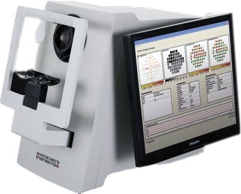
Heidelberg HEP Visual Field |
|
Early recognition of disease helps to delay and prevent the most common causes of blindness. |
Glaucoma is a group of eye diseases which result in damage to the optic nerve and vision loss. The most important examination in glaucoma patients is visual field assessment.
The Heidelberg Edge Perimeter (HEP) is an instrument for examining your visual field. Two different procedures can be used. One test is for early detection of glaucoma, and the other is for monitoring the progression of the disease.
|
|
|
Preparation
You will first be seated comfortably in front of the instrument and be handed a response button. During the entire examination, you will rest your chin on the instrument's chin rest. You will lean your forehead against the head rest so that you can keep your head still. During the visual field test, you will look at a monitor in the instrument and stare at a black dot in the middle of the screen. You can blink normally. If you need a break, the examiner can interrupt the test at any time and continue it a short time later. Your two eyes will be tested one at a time.
Examination I - Early glaucoma detection
The Heidelberg Edge Perimeter provides a flicker stimulus for early glaucoma detection. This stimulus is presented at irregular intervals, and you verify if you have perceived it. The instrument thus examines the so-called sensitivity of your retina, on which you can barely see the stimulus. When the examination begins, the instrument’s screen starts to flicker. A circle is illuminated for a short time at irregular intervals. The circles sometimes appear lighter, sometimes darker. Whenever you see a flickering circle, you press and release the response button. There are also phases when no stimulus is presented. Do not worry about it; that is completely normal.
Examination II - Monitoring progression in glaucoma
The examination with the white light stimulus is used for advanced cases of glaucoma and for monitoring of change in glaucoma over time. When the examination begins, a white point of light will appear on a gray background on the instrument’s monitor. The circles sometimes appear lighter, sometimes darker. Whenever you see a point of light, you press and release the response button.
Both methods determine your individual visual field. Deficiencies in the visual field can be
recognized on a printout. When visual field tests are repeated over several years, progression of deterioration can be documented and advancing glaucoma can be diagnosed, or the success of treatment can be measured.
This examination is painless and your vision will not be impaired. Afterwards, you can drive as usual, unless your
eye care professional has dilated your pupils to conduct a different examination. |
| |
|
| |
|
| |
|